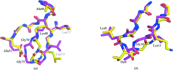
Figure 3.
Comparison of loop conformations in different Ub2 crystal structures. (a) Comparison of the isopeptide-bond conformation in the two Ub2 crystal structures. Chains A–B of the new crystal structure are coloured yellow and the previous structure (PDB code 1aar; Cook et al., 1992 ▶) is coloured magenta. Residues labelled with primes belong to a distal moiety. The conformation of the isopeptide bond in chains C–D and E–F is similar to that in chains A–B. (b) Comparison of the β1–β2 loop conformation in chain B (yellow) and the previous crystal structure (magenta). The conformation of this loop in chains C–D and E–F of the new structure is similar to that shown in magenta.

