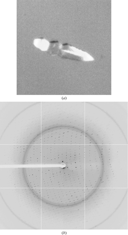
Figure 2.
Crystal and diffraction pattern of the MqsA–dsDNA complex used for data collection. (a) The MqsA–dsDNA-3 crystal used for data collection was grown in 0.2 M ammonium chloride, 20% PEG 3350 at 277 K in a drop containing 0.2 µl protein solution and 0.4 µl crystallization solution. (b) A representative diffraction image from the MqsA–dsDNA-3 crystal.

