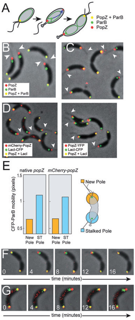
Fig. 2. The centromere tethering function of PopZ is lost at the stalked pole.
A. Schematic of ParB and PopZ localization during chromosome replication and centromere translocation. The chromosome is represented as a green oval, and the direction of movement during translocation is indicated by a blue arrow.
B. Cells of strain GB301, producing PopZ-YFP (red) and CFP-ParB (green). Fluorescence channels are overlayed on the phase-contrast image. Visible stalks are indicated by arrowheads. Fluorescence signals were considered ‘partially overlapping’ when 0–50% of the area of the smaller diameter signal was contained within the area of the larger diameter signal.
C. Cells of strain GB529, producing mCherry-PopZ (in red) and CFP-ParB (in green), processed as in (B).
D. In FROS (Straight et al., 1996; Viollier et al., 2004), exogenously expressed LacI-CFP binds to tandem arrays of lac operator sites inserted at a specific location on the chromosome, here a centromere proximal sequence. This labelling was performed in the context of mCherry-PopZ expression (left panel, strain GB593) and PopZ-YFP expression (right panel, strain GB594). Images were processed as in B, and visible stalks are marked by arrowheads.
E. Quantification of the motion of CFP-ParB foci at flagellated and stalked poles in time lapse movies of strain MT190 and GB529. Position was determined by calculating the weighted centroid of a fluorescent focus, and distance was measured in pixels, with a measured pixel width of approximately 80 nm. The plot shows average displacement after a four-minute interval. In three separate experiments, ~60 cells of each strain were analysed over four consecutive time intervals, yielding a total of over 700 data points. The standard error on the determination of the mean value was negligibly small.
F and G. Individual frames from time lapse movies of strain GB529 (F) and GB301 (G). Time (in minutes) is indicated. The CFP-ParB focus (green) moves back-and-forth in the vicinity of the stalked pole (top), but remains stationary at the flagellar pole (bottom). All experiments were performed in M2G medium.

