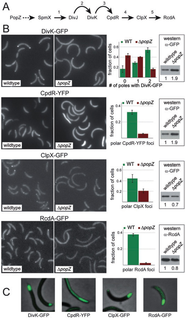
Fig. 5. PopZ mediates multi-protein complex assembly at the developing stalked pole.
A. An outline of the ClpXP protease localization and assembly pathway during stalked pole development. Curved arrows indicate control through phosphosignalling and straight arrows indicate physical protein interactions that are required for polar localization. The dotted line from PopZ to SpmX is inferred. References: 1: Radhakrishnan et al. (2008); 2: Jacobs et al. (2001); 3: Iniesta and Shapiro (2008); 4: Iniesta et al. (2006); 5: Mcgrath et al. (2006).
B. Localization of DivK-GFP, CpdR-YFP, ClpX-GFP and RcdA-GFP in wild-type and ΔpopZ cells grown in M2G medium. For each fluorescent protein, representative fluorescence image panels are placed beside quantified data from at least two independent experiments, each involving more than 200 cells, with error bars representing the standard deviation between trials. At the far right of each panel is a Western blot showing the cellular levels of the fluorescently tagged protein under these conditions. Below each blot, the numbers refer to the quantification and normalization of band intensities, with wild-type level set to 1. Reading from top left, the strains used were GB229, GB459, LS4250, GB460, LS4183, GB457, LS4191 and GB458.
C. Effects of PopZ overproduction on DivK-GFP, CpdR-YFP, ClpX-GFP and RcdA-GFP localization. Strains GB281, GB235, GB233 and GB265 were stimulated by growth in PYE medium supplemented with 0.3% xylose for eight hours prior to analysis.

