Figure 1.

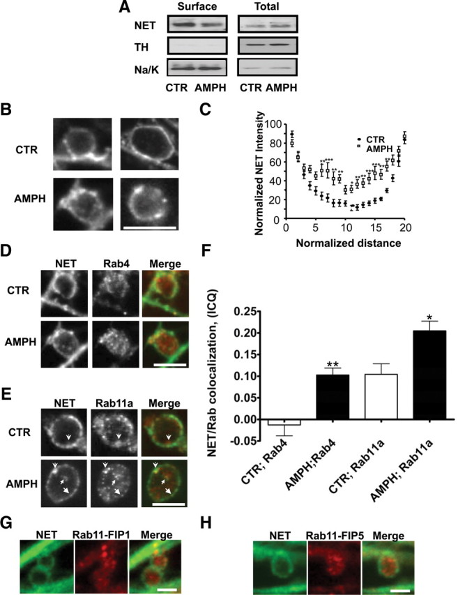
Amphetamine leads to a decrease in surface NET expression in rat cortical slices and NET in boutons and colocalization of NET with Rab11a and Rab4. A, Representative immunoblots of surface and total NET from biotinylated rat cortical slices. 300 μm rat cortical slices [paired right vs left hemispheres for vehicle (CTR) vs AMPH] were incubated for 30 min with 10 μm AMPH and then biotinylated (see Materials and Methods). A volume of 25 μl of total slice lysate was loaded per sample (lane). A volume of 45 μl of eluted (biotinylated proteins) was loaded per sample (lane). Tyrosine hydroxylase and Na/K ATPase immunoreactivity was also probed. A total of 13 cortical slices from three rats were analyzed. *p < 0.05 by Student's t test. Mouse SCGNs were cultured and processed for immunocytochemistry as described in Materials and Methods. B, SCGNs treated with 10 μm AMPH for 30 min leads to an accumulation of intracellular NET in SCGN boutons. Images are taken from single confocal and depict single boutons with associated axons (linear structures). The perimeter of the boutons is marked by intense NET immunoreactivity, as well as intrabouton immunoreactivity, which is increased by AMPH. C, Analysis of intensity plots spanning SCGN boutons demonstrates that AMPH treatment induces NET accumulation in the interior of these boutons. The normalized NET intensity is plotted against the normalized distance as described in Materials and Methods. D, AMPH treatment of SCGNs enhances NET and Rab4 colocalization in boutons. Shown in the top row are boutons from cultures treated with vehicle (basal state). Shown in the bottom row are AMPH-stimulated cells showing colocalization between NET and Rab4. E, AMPH treatment of SCGNs enhances NET and Rab11a colocalization in boutons. Shown in the top row are boutons from cultures treated with vehicle (basal state), and arrowheads indicate colocalization of NET and Rab11a at a juxtaplasma membrane location. The intensity of the juxtaplasma membrane NET is lower than on the perimeter. Shown in the bottom row are AMPH-stimulated cells with arrows pointing to surface (arrows), juxtaplasma membrane (arrowhead), and internal (small arrow) regions showing colocalization between NET and Rab11a. F, Quantitation of NET and Rab11a, Rab4 colocalization using the ICQ as outlined in Materials and Methods (Li et al., 2004) demonstrates that AMPH enhances the colocalization of NET and Rab11a as well as Rab4. G, H, Rab11a effectors are present in the presynaptic compartments of SCGN. Scale bar, 5 μm. Error bars indicate SEM.
