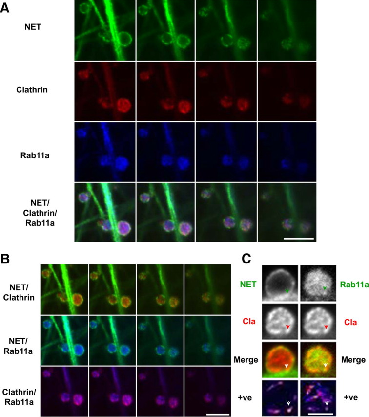
Figure 4.

At early time points of AMPH treatment of SCGNs, Rab11a colocalizes with clathrin at juxtaplasma membrane and internal sites. Mouse SCGNs were cultured, treated with vehicle or AMPH (10 μm) for 10 min, and processed for immunocytochemistry as described in Materials and Methods. All images are taken from single confocal sections. Cultures were treated with vehicle or AMPH (10 μm) for 10 min. At this time point, low levels of NET are internalized (Fig. 2). A, In this panel, single confocal sections spanning the boutons from the center to the bottom are displayed. They are tripled labeled for NET, clathrin, and Rab11a. The merge of the triple-labeled sections is shown in the fourth row. Scale bar, 2.5 μm. B, Displayed are the double-merge images of the same sections as in A. Some colocalization of clathrin with NET is detected at the edges of NET puncta (top row), and low levels of NET and Rab11a can be seen at the perimeter (middle row). Finally, clathrin and Rab11a are highly colocalized both at the surface and in the interior (bottom row). Scale bar, 2.5 μm. C, In this panel, single confocal sections at the center to the bottom are displayed. This section was tripled labeled for NET (NET), clathrin (Cla), and Rab11a (Rab11a). For the sake of comparison, the double-merged image was generated by pseudocoloring both NET and Rab11a red, whereas clathrin was green. In the bottom row are images that display pseudocolored pixels from the images of boutons in which the pixel values for the two relevant antigens are both above the mean. An arrowhead indicate a site in which both NET/clathrin colocalize and Rab11a/clathrin colocalize based on analyzing the two images in the bottom panel (see Materials and Methods). Scale bar, 2.5 μm.
