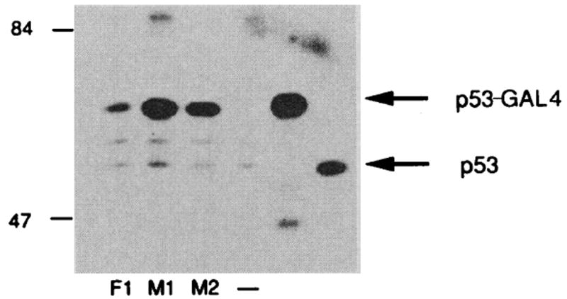
Fig. 4.

Immunoprecipitation of p53-GAL4 fusion proteins. Plasmids encoding wild-type p53-GAL4 (Fl), p53-GAL4 mutant 1 (Ml), p53-GAL4 mutant 2 (M2), or vector alone (–) were cotransfected with a plasmid containing the β-galactosidase gene into COS cells with DEAE dextran. The β-galactosidase plasmid was used to monitor transfection efficiency. Transfected cells were labeled with a mixture of 35S-labeled Cys and 35S-labeled Met. Immunoprecipitations were performed as described with p53 monoclonal antibody PAb248 and equal amounts of trichloroacetic acid–precipitable counts (28). Also shown are in vitro–translated wild-type p53- GAL4 fusion 1 (lane 5) and p53 mutant 1 immunoprecipitated from cells that overexpress this protein (lane 6). Molecular weight markers are shown in kilodaltons.
