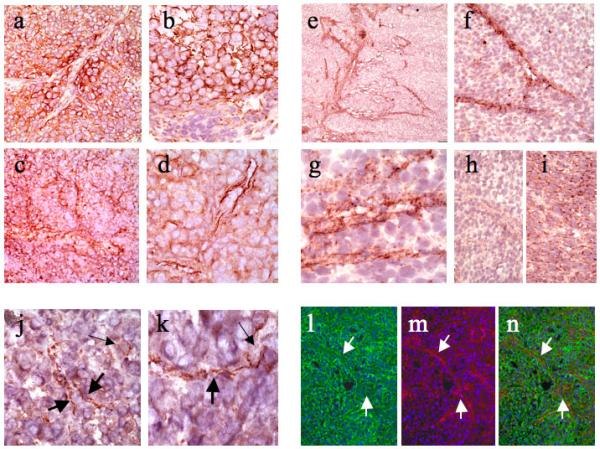
Figure 2. Nodal Protein and mRNA Expression in Humanized Xenografts and Patient-Derived Melanomas.

(a and b) Immunohistochemistry for Nodal protein revealed focally diffuse patterns of reactivity localized to melanoma cell cytoplasm and cell membranes. Note in Figure 2a, however, that in the center of the field Nodal stains channel-like structures. (c and d) In some fields, Nodal reactivity is accentuated in a spatial distribution suggestive of patterned networks of channel formation. (e and f) In situ hybridization for Nodal mRNA disclosed striking localization to anastomosing networks spatially identical to those forming VM channels. (g) High-power view of Nodal mRNA expression in a network forming lumen-like spaces consistent with true channel formation. (h) Negative control with Nodal RNA sense probe. (i) Positive control with housekeeping genes. (j and k) Nodal staining of patient-derived melanomas highlighted similar delicate intratumoral channels. (l) Immunofluorescence with Nodal (in green) showed localization to cell membranes and a channel-like pattern. (m) Immunofluorescence with VE-Cadherin (in red) highlighted VM channels. (n) Superimposed images showed colocalization (in yellow) of Nodal and VE-cadherin in the VM channels.
