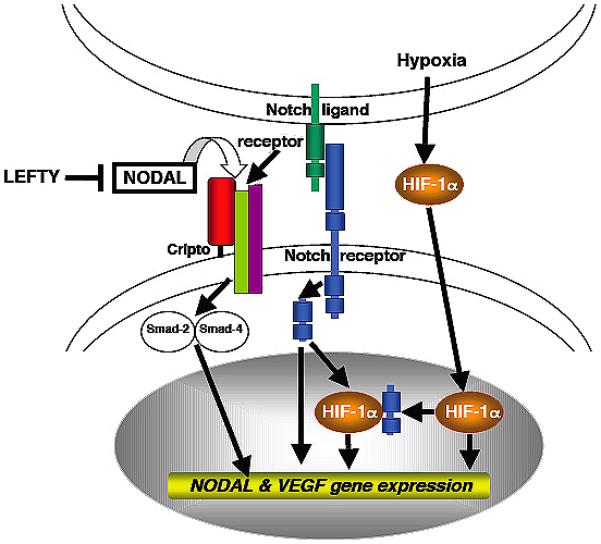
Figure 3. Proposed Nodal signaling pathway in vasculogenic mimicry.

Rapid growth of early melanoma tumor nodules causes them to outgrow their blood supply and creates hypoxia. This stimulates expression of hypoxia-induced factor 1-α (HIF-1α), which activates VEGF. VEGF induces the formation of VM networks. Concurrently, HIF-1α signals to the intracellular domain of Notch. Once activated, the intracellular component of Notch translocates to the nucleus and activates transcription of Nodal. Nodal is then secreted, where it induces Nodal expression by melanoma cells. Melanoma cells signal to one another in an autocrine manner via Nodal, maintaining a stem cell-like state capable of continued endothelial differential and VM formation.
