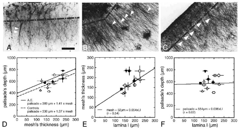
Fig. 1.

Astroglial architectures in the brain of AE and control cases: presence of the “interlaminar palisade” and of stellate (intralaminar) astrocytes (mostly in lamina I). (A–C) (A) Case AE, occipital cortex, block #185; (B) case H59, frontal cortex, area 8/46; (C) case H60, occipital cortex, area 17/18. Note periodic aggregates of interlaminar processes (single arrowheads) (B, C), and occasional fascicles (double arrowheads) within it (A). Broken line indicates extent of lamina I. Bar (A–C): 100 μm. (D) Linear regression performed on AE (continuous line) and control cases (dashed line) shows a common trend of data points in all samples. Also, the superficial glial net and the thickness of lamina I showed a good correspondence (E). On the contrary, no relation was found between the length of interlaminar processes and the thickness of lamina I (F). Analyzed regions: prefrontal cortex (Brodmann's) area 8/46 (triangle facing up); occipital cortex, area 17/18 and block #185 (AE) (square); frontal cortex, block #211 (AE) (diamond); inferior parietal cortex, block #106 (AE) (circle); parietal somatosensory cortex, block #49 (AE) (triangle facing down).
