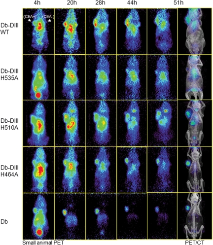
Fig. 4.
Small animal PET/CT imaging of athymic nude mice xenografted with CEA-positive LS174T (left) and CEA-negative C6 (right) tumors. Groups of four mice were injected with 124I-labeled Db-DIII proteins (WT, H535A, H510A or H464A) and the anti-CEA Db as a reference. Mice were imaged for 10 min at five different time points with coronal sections shown. Co-registered PET/CT images are included for the anatomical reference of the tumors and organs.

