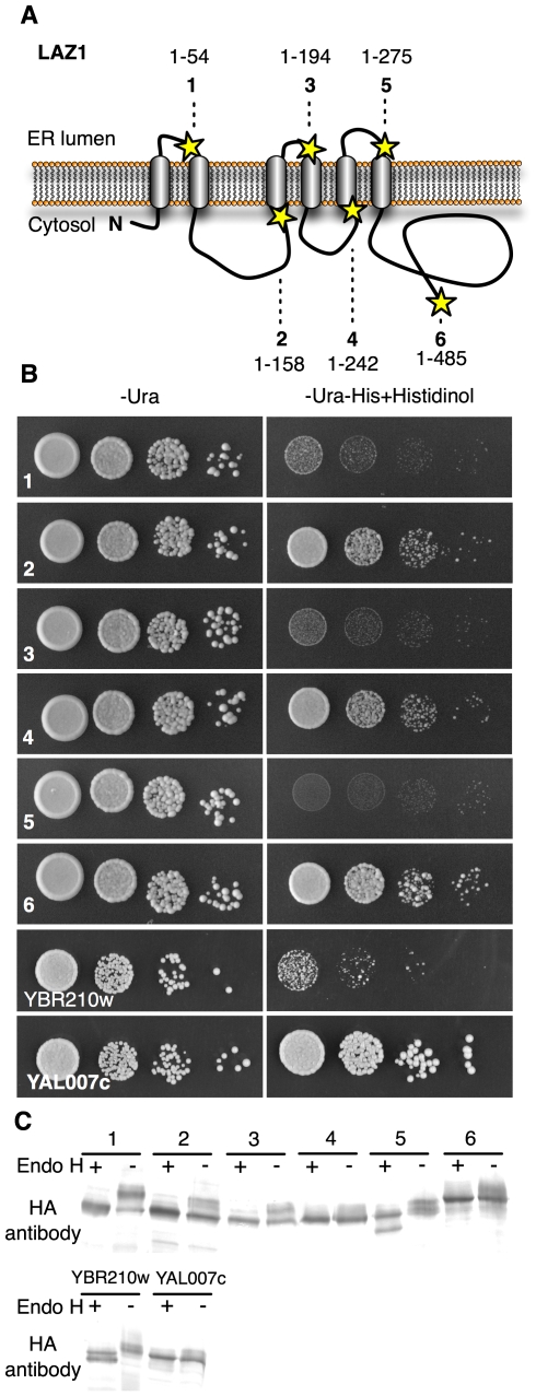
Figure 5. LAZ1 topology.
(A) Six LAZ1 forms (1–6) were C-terminally fused to the dual SUC2/HIS4C reporter to determine their orientation in membranes. These LAZ1 fusions were chosen to assess the validity of predicted transmembrane regions (Figure S4). Lengths and positions of the fusions are indicated by lines and stars, respectively. (B) Growth of yeast harboring the six LAZ1 reporter fusions on histidinol containing medium indicates cytosolic localization of the reporter due to its histidinol dehydrogenase activity. YBR210w (ER localized C-terminus) and YAL007c (cytosolic localized C-terminus) served as controls [24]. (C) The glycosylation status of six LAZ1 fusions (Fig. 5A) was determined by comparing the sizes of endoglycosidase H (endo H) treated and untreated samples followed by Western blotting using anti-HA antibody. Faster migrating (lower) bands after endo H treatment indicate that the fusion is glycosylated and the SUC2/HIS3 C-terminal reporter resides in the ER. YBR210w (ER localized C-terminus) and YAL007c (cytosolic localized C-terminus) served as controls [24].

