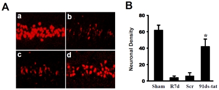
Figure 4. Contributions of NADPH oxidase to Ischemic Neuronal Damage in Hippocampal CA1.
(A) Representative staining of coronal CA1 sections with NeuN (red) show neuroprotection of gp91ds-tat peptide at day 7 after reperfusion. a: Sham; b: Ischemic reperfusion; c: Scrambled gp91ds-tat control; d: gp91ds-tat (91ds-tat). (B) CA1 neuronal density was counted per 250 µm length of medial CA1 region from five to six rats in each group. *p<0.05 vs. reperfusion at 7 days (R7d) and Scr treatment groups. Scr: gp91ds-tat control.

