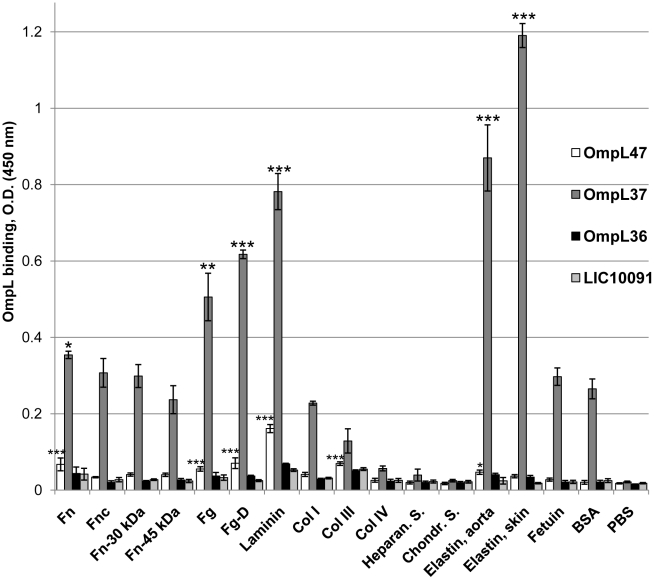
Figure 1. Binding of recombinant Omp36, OmpL37, and OmpL47 to host tissue components.
Microtiter wells were coated with 1 µg of plasma fibronectin (Fn), fibroblast cellular fibronectin (Fnc), heparin-binding domain of plasma fibronectin (Fn-30 kDa), gelatin-binding domain of plasma fibronectin (Fn-45 kDa), plasma fibrinogen (Fg), plasma fibrinogen fragment D (Fg-D), laminin (Lm), collagen type I (Col I), collagen type III (Col III), collagen type IV (Col IV), kidney heparan sulfate (Heparan. S.), cartilage chondroitin sulfate (Chondr. S.), aorta elastin, skin elastin, fetuin, and BSA. One microgram of recombinant protein was added per well and binding was measured by ELISA. Data represent the mean absorbance at 450 nm ± the standard deviation of three independent experiments. The binding of recombinant proteins to tissue components was compared to their binding to BSA by Student's two-tailed t test (*** P<0.001, ** P<0.01, * P<0.05).

