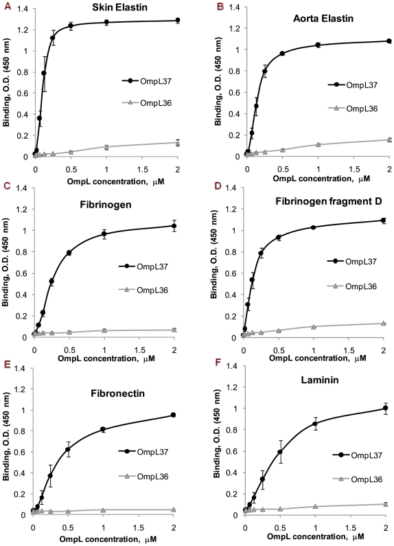
Figure 2. Binding to skin and aorta elastin, fibrinogen, fibrinogen fragment D, fibronectin, and laminin as a function of OmpL37 concentration.
Binding of OmpL37 (concentration ranging from 0 to 2 µM) to 1 µg of immobilized (A) human skin elastin, (B) human aorta elastin, (C) human plasma fibrinogen, (D) human plasma fibrinogen fragment D, (E) human plasma fibronectin, and (F) murine laminin was measured by ELISA. The mean optical density at 450 nm ± the standard deviation of three independent experiments is shown at each point. The apparent Kd for saturating binding was estimated as the concentration of recombinant OmpL37 resulting in half-maximal binding (see text and Table 1). OmpL36 served as a negative control.

