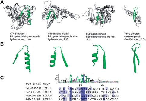
Fig. 2.
Matches of the nucleotide–triphosphate-binding (p-loop) prototype in crystal structures. Four matches of nucleotide–triphosphate-binding prototype are shown. The fold is displayed in cartoon and the structural loop corresponding to p-loop prototype is highlighted in green. The structures of the EFLs are also displayed. The logo of the prototype and the alignment of sequences of the corresponding EFLs highlight the functionally important residues involved in nucleotide–triphosphate binding and hydrolysis. PDB ID, SCOP ID and the coordinates of the sequence segments corresponding to the domains which contain the EFLs on display, are shown in the bottom.

