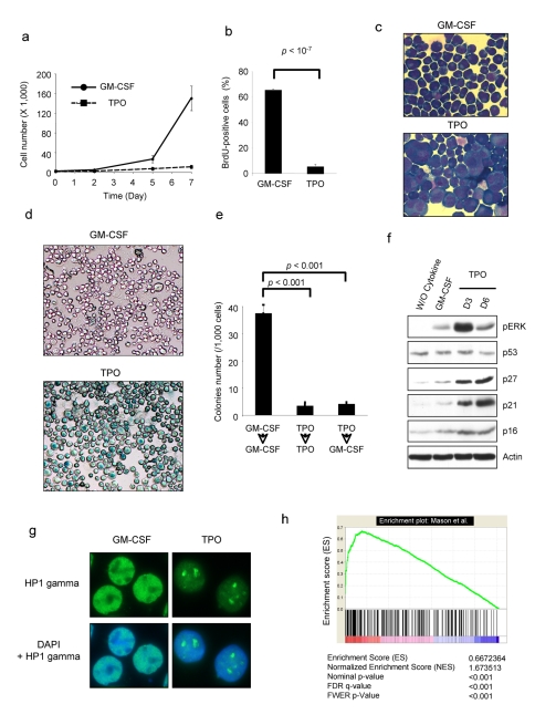
Figure 1. Thrombopoietin induces cellular senescence of UT711oc1 cells.
(a) TPO inhibits UT711oc1 cell proliferation. UT711oc1 cells were cultured in presence of either GM-CSF or TPO. Viable cells were counted using Trypan blue exclusion. (b) TPO induces a decrease in DNA replication in UT711oc1 cells. BrdU incorporation was measured in UT711oc1 exposed for 5 d to either GM-CSF or TPO. (c) Morphological changes in TPO-stimulated UT711oc1 cells. Cells were grown for 5 d in presence of either GM-CSF or TPO and stained with May-Grünwald Giemsa. (d) SA-β-galactosidase staining in TPO-treated cells. (e) Irreversible cell-cycle arrest. UT711oc1 cells were grown for 5 d with GM-CSF or TPO and seeded in methylcellulose with GM-CSF or TPO. Cell colony numbers were determined. (f) Sustained ERK phosphorylation and p21 expression after TPO exposure. UT711oc1 cells were cultured in presence of GM-CSF or TPO and proteins were analyzed by Western blotting. (g) Formation of heterochromatin foci in UT711oc1 cells treated with TPO. Cells were grown with either GM-CSF or TPO and heterochromatin protein 1 (HP1) gamma was revealed by immunofluorescence. (h) GSEA. After 12 h cytokine starvation, UT711oc1 cells were stimulated with either GM-CSF or TPO. TPO-induced gene expression—relative to GM-CSF—was determined by micro-array analysis and compared by a Gene Set Enrichment Analysis with the molecular signature of oncogenic ras-induced senescence determined in fibroblasts by Mason et al. [29]. In histograms shown, error bars represent standard deviations of three independent experiments.

