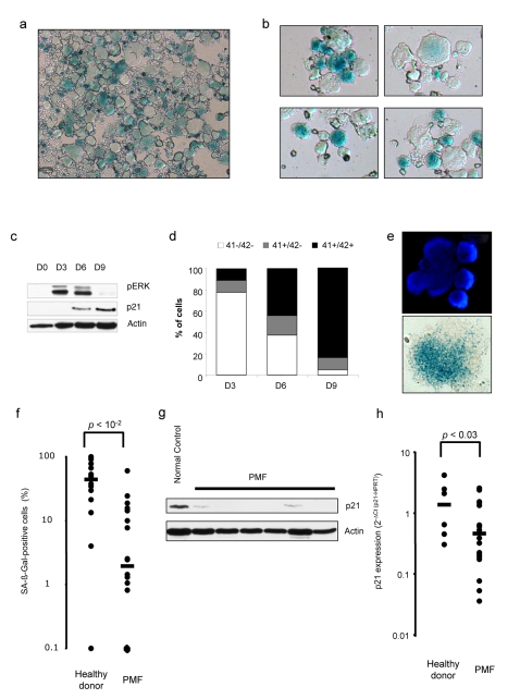
Figure 5. Cellular senescence is present in human and mouse mature megacaryocytes but is lacking in oncogenic megakaryocytes.
(a) C57/Bl6 purified Lin− were cultured in serum-free medium with 10 ng/mL TPO for megakaryocytic differentiation and stained at day 6 to reveal a SA-β-galactosidase activity. (b) Human CD34+ cells were cultured in serum-free medium for 10 d and SA-β-galactosidase activity was detected. (c) ERK phosphorylation status and p21 expression were analyzed by Western blotting during human megakaryopoiesis at days 0, 3, 6, and 9. (d) Levels of megakaryocyte maturation membrane markers (CD41+ and CD42+) increase during the culture of CD34+ cells into megacaryocytes. (e) Human megakaryocytes isolated from healthy bone marrow revealed a SA-β-galactosidase activity. One cell isolated from several present in the original image is represented. (f) Percentage of SA-β-galactosidase-positive megakaryocytes per sample were analyzed in normal and primary myelofibrosis megakaryocytes (PMFs). PMFs compared to normal megakaryocytes in culture show a significant decrease in (g) p21 protein expression and (h) p21 mRNA level. Scale bar indicates the sample median. Error bar represents the standard deviation. Each dot represents one PMF or healthy donor sample.

