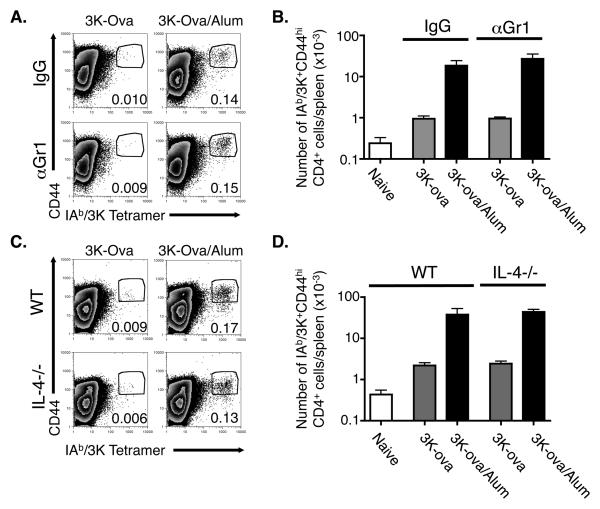
Figure 5. The ability of alum to enhance expansion of antigen specific CD4 cells is independent of Gr1 expressing cells and IL-4.
A,B. C57BL/6 mice were injected with 3K-Ova either alone or adsorbed to alum and treated with either rat IgG or anti-Gr1 as described in Materials and Methods. Nine days later splenocytes were stained as in Figure 4. A. The FACS plots show the staining of F4/80- B220- CD4+ cells with anti-CD44 and IAb/3K tetramer. Numbers indicate the percentage of F4/80- B220- CD4+ cells that were CD44hi and IAb/3K tetramer positive and are representative of three individual samples. B. Shown are the average +/− standard error of the numbers of CD44hi IAb/3K-staining CD4+ cells/spleen of mice immunized as indicated. Results are representative of three independent experiments. C.D. Wild type or IL-4−/− mice were injected with 3K-Ova with or without alum. Nine days later their spleen cells were isolated and analyzed as in A, B. Results are representative of two separate experiments.

