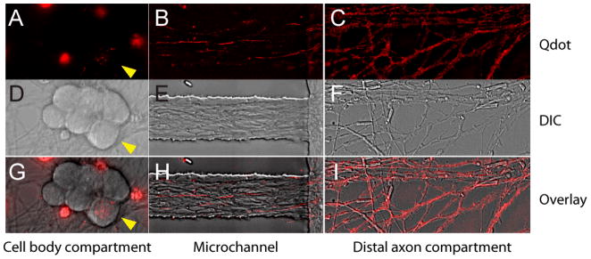
FIGURE 3.
Image of axons containing Qdot-NGF in different compartments of the microfluidic device (three-compartment design). Fluorescent Qdot-NGF (1 nM) was added to the distal axon compartment and incubated for 2hrs prior to imaging. A–C) Fluorescence images of Qdot-NGF in (A) cell-body compartment, (B) microchannel neighboring the cell body compartment and (C) distal-axon compartment. The image in (B) is the projection of 600 time-lapse frames, so that the traces of individual Qdot-NGF depict axons that they traveled along. D–F) DIC images of the same areas as (A–C). G–I) overlaid images of fluorescence and DIC images in each compartment. Note that only one DRG cell body (yellow arrow heads) out of six cell bodies contains Qdot-NGFs. This is because not all neurons were able to extend their axons into the distal axon compartment, which further confirms the liquid isolation between the cell body compartment and the distal axon compartment. The large fluorescence splotch in (A) and (G) arises from the autofluorescent cell debris.

