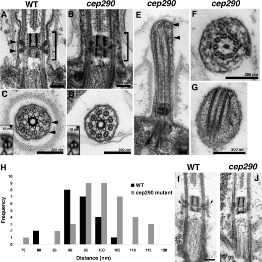
Figure 3.
Loss of CEP290 causes defects in the microtubule–membrane connections within the transition zone. (A and B) Longitudinal sections through the transition zone (brackets) reveal that the electron-dense wedge-shaped structures (arrowheads in A) between the transition zone microtubules and flagellar membrane are missing in the cep290 mutant. (C and D) Cross sections reveal that the Y-shaped connectors (arrowheads in C) that bridge the transition zone microtubules with the membrane are missing from most cep290 doublet microtubules. Insets in C and D are image averages of 30 transition zone doublet microtubules. (E–G) Some of the mutant flagella have bulges filled with electron-dense material (arrowheads in E). The flagellum shown in F also has defects in axonemal doublets and the central pair microtubules; however, such defects are unusual in the mutant, as most axonemal cross sections appear normal. (H) Histogram depicting the distribution of the distances between the transition zone cylinder and the flagellar membrane in wild-type and cep290 mutant cells; the difference in the distributions is significant at P = 0.0025 for a Student’s t test and P = 0.0009 for a Welch’s t test. (I and J) Cells were detergent-extracted, then fixed and processed for EM. The detergent-resistant membrane that remains attached to the wild-type transition zone (arrows in I) is no longer attached to the mutant transition zone. Bar, 100 nm.

