Figure 2.
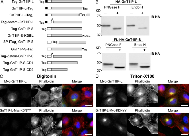
GnT1IP-L is a type II transmembrane glycoprotein with high mannose N-glycans. (A) GnT1IP constructs showing the N-terminal cytosolic domain of GnT1IP-L (aa 1–48; black box); the predicted transmembrane domain of GnT1IP-L (aa 49–69; white box) or signal peptide (SP) of GnT1IP-S (aa 1–26); the Golgi lumenal domain (aa 70–417 for GnT1IP-L and aa 27–373 for GnT1IP-S; gray box), and the C-terminal deletion mutants Tag-GnT1IP-S-CD1 (39 aa deletion) and -CD2 (122 aa deletion); Tag represents FLAG-HA (FL-HA), HA, or Myc; internal tags were inserted after aa 412 of GnT1IP-L (iTag1) and after aa 26 of GnT1IP-S (iTag2); the 48 aa stem-region deletion (Δ stem) from aa 71–118 in GnT1IP-L and aa 27–74 in GnT1IP-S is shown by a hat. The KDEL sequence was inserted after aa 373 of GnT1IP-S. (B) Lysates from CHO cells expressing HA-GnT1IP-L or FL-HA-GnT1IP-S digested with PNGase F or Endo H (+) or incubated without enzyme (−) and subjected to immunoblotting using anti-HA mAb (IB HA). (C) HeLa cells transiently expressing Myc-GnT1IP-L or GnT1IP-L-Myc-KDNYY were fixed, treated with 5 µg/ml digitonin or (D) 0.2% Triton X-100, immunolabeled for Myc-tagged GnT1IP-L (green) and actin (phalloidin; red), and observed by fluorescence microscopy. Bars, 20 µm.

