Figure 1.
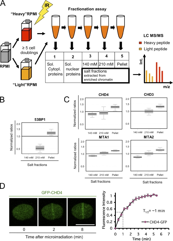
Identification of CHD4 as a factor involved in the DDR. (A) Proteomic screening procedure. GM00130 lymphocytes were grown in heavy or light SILAC media, exposed to 10 Gy of IR, fractionated, and analyzed by tandem mass spectrometry (MS/MS). LC, liquid chromatography. (B) Box plot showing quantitative tandem mass spectrometry data for 53BP1 (positive control). Y axis, normalized ratios (IR peptide/control peptide) showing protein elution by progressive salt fractionation of irradiated lymphocytes relative to control lymphocytes. The box represents the central 50% of the distributions, and the whiskers approximate the 95% interval. (C) Tandem mass spectrometry data for NuRD subunits are shown. Box plots are as in B. (D) Accumulation of GFP-CHD4 at laser-generated DSBs (left) and real-time recruitment of GFP-CHD4 derived from 10 independent cells (right). Error bars indicate SEM. Bar, 10 µm.

