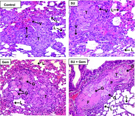
Figure 3.
Histology of spontaneous lung metastases from mice treated with sunitinib and gemcitabine. Lungs from mice treated with sunitinib (20 mg/kg), gemcitabine (20 mg/kg), and both combined, obtained on day 28 from experiments described in Figure 1, were processed for histology and H&E staining. The main findings were labeled on the prints with T for tumor, V for vessels, G for giant tumor cells, F for fibrotic areas, and L for normal lung alveoli. Metastastic lung tumor nodules from untreated mice (Control) consisted of tumor cells with pleomorphic nuclei and prominent nucleoli and contained areas of dilated vessels. Sunitinib (SU)-treated mice had decreased number of tumor cells and vessels in lung tumor nodules. Lung tumor nodules from gemcitabine (Gem)- treated mice showed giant tumor cells with cytoplasmic vacuoles and eosinophilic inclusions and decreased vascularization. Gemcitabine combined with sunitinib (SU + Gem) contained large eosinophilic areas of hyalinization, fibrosis, and a few giant abnormal tumor cells. Figures were enlarged to show changes in lung tumor nodules.

