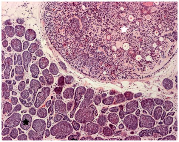
Figure 4.
Haematoxylin and eosin stain displaying the characteristic pattern suggestive of cylinders in cross section which gave rise to the term cylindroma (lower left corner; black star), with an adjacent region (upper right corner; white star) within the tumor displaying a large ball of basophillic cells with areas of ductal differentiation, consistent with an eccrine spiradenoma (Original magnification 10x).

