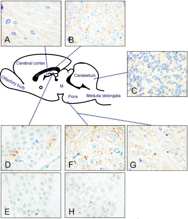
Figure 3.
Immunohistochemical staining of CA XIV in mouse brain. The schematic model indicates approximate anatomical sites which are shown in A–H. Strong positive staining by the biotin–streptavidin complex method, with anti-mouse CA XIV serum as first antibody, is seen in the large neuronal bodies and axons located in the basal part of pons (F and G). Other CA XIV-positive sites shown in this figure include corpus callosum (A), hippocampus (B), granular cell layer of cerebellum (C), and choroid plexus (D). The control sections of choroid plexus (E) and pons (H), with preimmune serum as first antibody, were negative. Original magnifications: A, G, and H, ×400; B and F, ×500; C, D, and E, ×250. CC, corpus callosum; H, hippocampus; M, midbrain.

