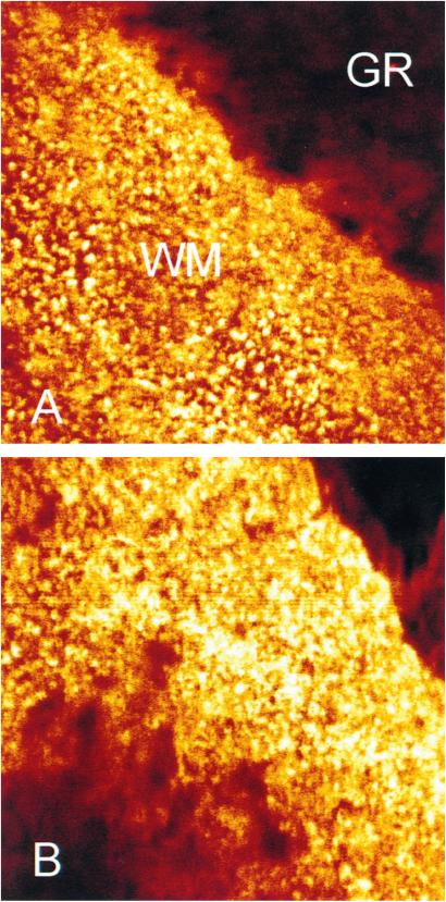
Figure 4.
Confocal laser scanning microscopy images of immunofluorescently labeled CA XIV in frozen sections of mouse brain. Punctate staining is located in the white matter (WM) of cerebellum (A) and anterolateral part of pons (B). The granular cell layer of cerebellum (GR) shows only a weak positive reaction. Original magnification: ×630.

