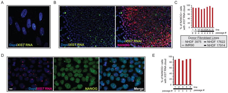
Figure 2. Female hiPSCs carry a single XIST RNA- coated chromosome.
(A) FISH for XIST RNA (green) in the Dapi-stained fibroblast line NHDF 17430. The accumulation of XIST RNA on the Xi can be seen as focal nuclear enrichment of the RNA signal.
(B) FISH for XIST RNA (green) in the STEMCCA-hiPSC line E, merged with Dapi in the left image and overlaid with an immunostaining for NANOG (red) in the right image.
(C) The graph depicts the percentage of NANOG-positive cells with a single Xi-like accumulation of XIST RNA for various STEMCCA-hiPSC lines. Fibroblast origin is marked with gray scale indicators and passage of hiPSCs is indicated.
(D) As in (B) with NANOG in green and XIST RNA in red for MIP-hiPSC line 1. Scale bar denotes 10um.
(E) As in (C) for various MIP-hiPSC lines. See Fig S2 for additional data.

