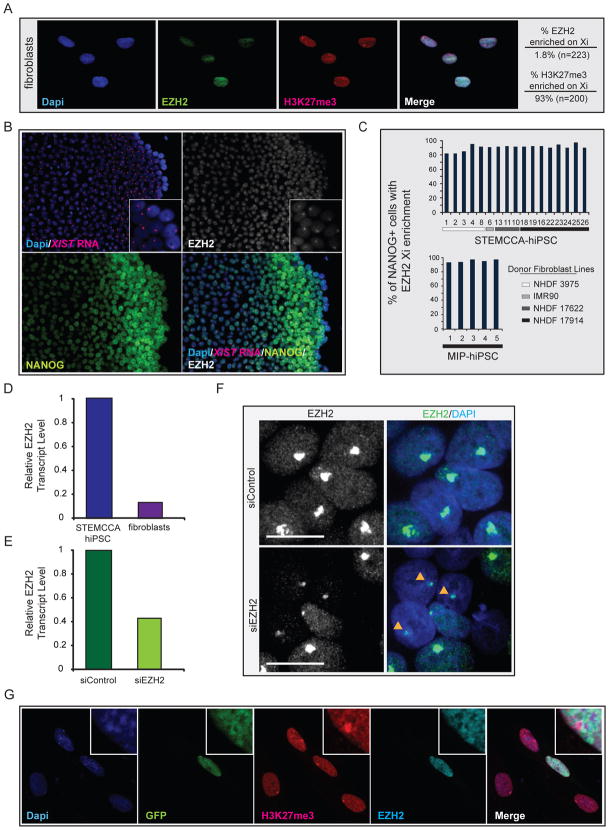
Figure 5. Characterization of Xi chromatin in female hiPSCs.
(A) Immunostaining for EZH2 (green) and H3K27me3 (red) in NHDF17914 fibroblasts, nuclei were detected with Dapi. The proportion of cells with an Xi-like accumulation for H3K27me3 and EZH2 are given.
(B) Immunostaining for EZH2 (grey, from far-red channel), NANOG (green) and FISH for XIST RNA (red) in STEMCCA-hiPSC line G at passage 8.
(C) Proportion of NANOG-positive cells with an Xi-like accumulation of EZH2 for various MIP and STEMCCA-hiPSC lines at around passages 4–8. See Fig S5 for additional data.
(D) Real-time PCR data for EZH2 transcript levels in given cell types, normalized to GAPDH.
(E) Real-time PCR data for EZH2 transcript levels in early passage STEMCCA-hiPSCs upon treatment with scrambled siRNA controls (siControl) or siRNAs targeting EZH2 (siEZH2), normalized to GAPDH.
(F) Immunostaining for EZH2 in an early passage STEMCCA-hiPSC line upon transfection of control or EZH2 siRNAs. Cells are mosaic for depletion of EZH2 and arrowheads point to EZH2 Xi enrichment in cells with clear EZH2 knockdown.
(G) Immunostaining for histone H3K27me3 (red) and EZH2 (far-red, pseudocolored in cyan) in fibroblasts two days after transfection of a GFP-EZH2 overexpression plasmid. The zoom in suggests that higher global levels of EZH2 in fibroblasts do not lead to Xi-enrichment of EZH2.

