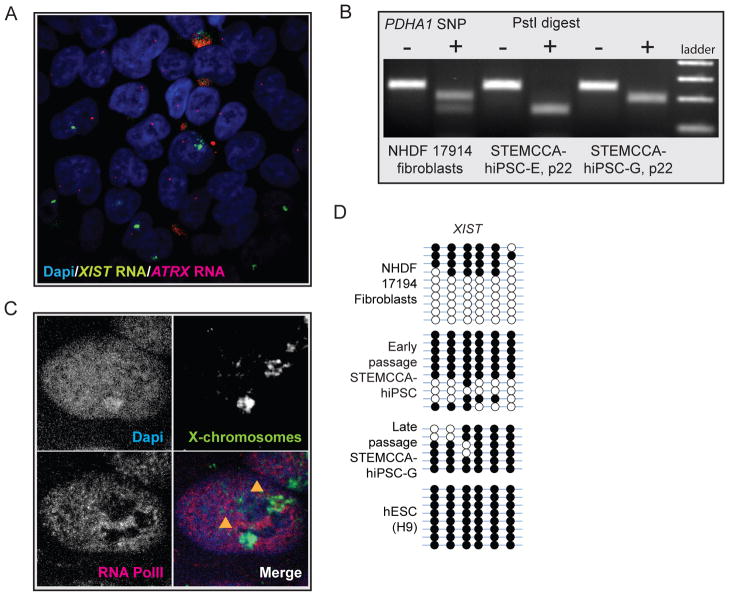
Figure 6. hiPSC lines are prone to XIST loss upon extended passaging but maintain the inactive state.
(A) RNA FISH for XIST (green) and ATRX (red) transcripts in STEMCCA-iPS line G at passage 16. At this passage, many cells without XIST RNA clouds were found. Note that ATRX continues to be expressed from only one chromosome. At around passage 22, this hiPSC line lost XIST RNA expression and coating in almost all cells (data not shown).
(B) Analysis of allele-specific expression of PDHA1 in STEMCCA-hiPSC lines G and E at passage 22 (upon loss of XIST) using our SNP-based assay. NHDF17914 fibroblast analysis was added as reference for a cell line expressing both alleles of PDHA1. Upon XIST loss, both hiPSC lines continue to express only the PHDA1 allele that was active at earlier passage (compare with Fig 4D) and do not reactivate the other allele.
(C) X chromosome paint (green) in STEMCCA-hiPSC lines G at passage 22 to detect the presence of two X chromosomes. The combination of the X-paint with RNA polymerase II immunostaining (red) illustrated that one of the two X chromosomes labeled with arrowheads is completely excluded from RNA polymerase II staining indicative of an Xi.
(D) Bisulfite sequencing of promoter region of XIST in indicated cell lines. Half of the clones were completely methylated in female fibroblasts and early passage STEMCCA-hiPSC in agreement with the notion that XIST is silenced on the Xa and actively expressed from the Xi. Complete methylation was found in STEMCCA-hiPSC line G at passage 22, which has lost XIST expression, and our H9 hESC culture, which also lacks XIST expression but contains an Xi (data not shown).

