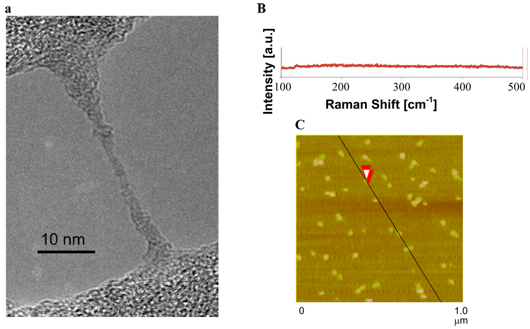
Figure 1.
(a) TEM image of HCCs spanning an empty domain on a lacy carbon grid. The HCCs have aggregated during the sample preparation, which consisted of placing an aqueous dispersion of HCCs on the lacy carbon grid and drying the sample at 70 °C for 16 h. (b) Raman spectroscopy indicates that the HCCs have very few or no radial breathing modes. (c) AFM image of aggregated HCCs on mica. The overlapping red arrows indicate where the height the HCCs was measured to be 1.9 nm.

