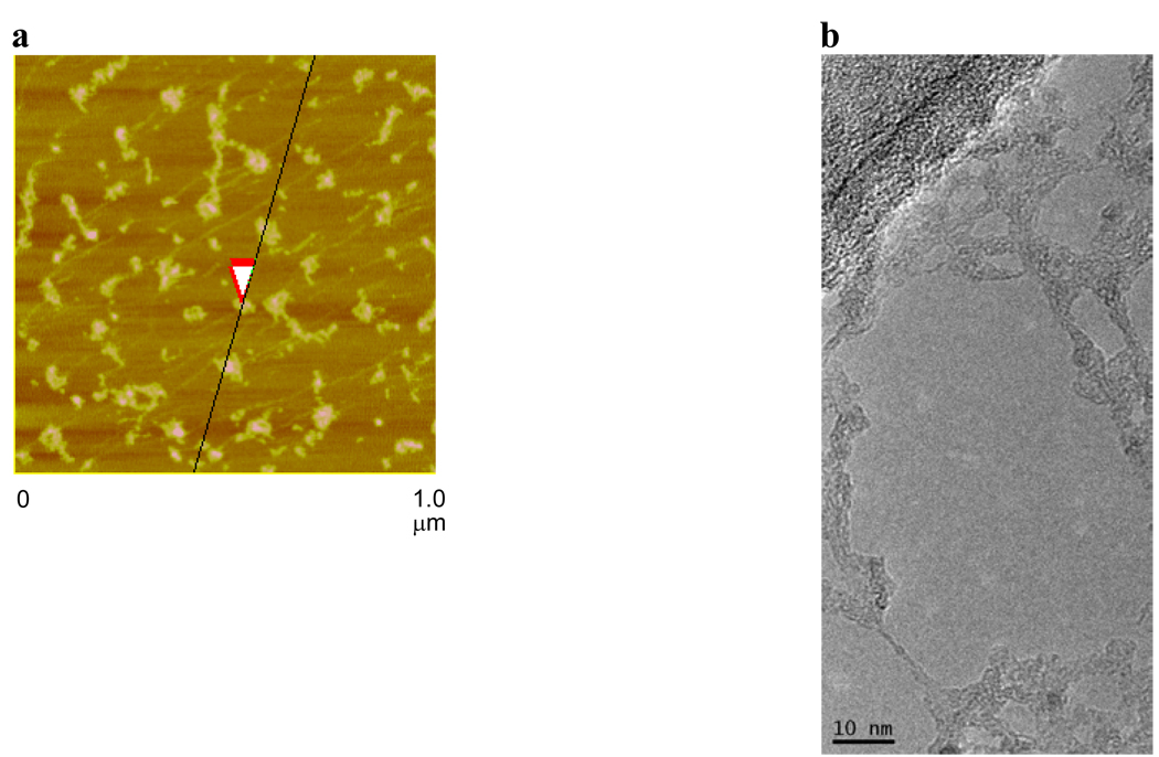
Figure 2.
(a) AFM image of aggregated PEG-HCCs on mica. The overlapping red arrows indicate where the height of the PEG-HCCs was measured to be 1.7 nm. (b) TEM image of PEG-HCCs spanning an empty domain on a lacy carbon grid. The PEG-HCCs have aggregated during the sample preparation, which consisted of placing an aqueous solution of PEG-HCCs on the lacy carbon grid and drying the sample at 70 °C for 16 h.

