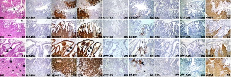
Figure 1.
CT antigen expression in week 5 placenta. (A) Overview, (B) villi with placenta bed, (C) villi, (D) placenta bed with invasive trophoblast. Hematoxylin-eosin staining (A1, B1, C1, D1). (A1) Overview with centrally located amnion cavity (*) and circumferential placental parenchyma. (B1) Magnified area of (A1) showing full thickness of placental parenchyma from amnion cavity (*) to endometrium (**) with invasive trophoblast. (C1) Placental villi with trophoblast cell column (arrows). (D1) Endometrium with broad band of the invasive trophoblast (+) with several extravillous syncytiotrophoblastic cells (thin arrows) close to the tips of villi (thick arrows). Negative immunohistochemical staining with mAb MA454 to MAGE-A1 (A2, B2, C2, D2), mAb CT7-33 to CT7/MAGE-C1 (A5, B5, C5, D5), mAb #23 to GAGE (A7, B7, C7, D7), and CT10#5 to CT10/MAGE-C2 (A8, B8, C8, D8). Homogeneous immunoreactivity of mAb M3H67 (A3, B3, C3, D3) and mAb 57 (A4, B4, C4, D4) in two distinct areas (A3, A4) comprising villous and extravillous trophoblast (B3, B4). Intense immunoreactivity of villous cytotrophoblast (thick arrows) and syncytiotrophoblast (thin arrows), as well as extravillous trophoblast of cell columns (*) (C3, C4), is observed. (D3, D4) Intensely stained deeper areas of the invasive extravillous trophoblast. Focal immunoreactivity of mAb ES121 to NY-ESO-1 (A6), involving primarily the villi (B6) with immunolabeling of the villous cytotrophoblast (thin arrows) (C6) and smaller foci of a cell column (thick arrow) with immunonegative invasive extravillous trophoblast (*)(D6). Homogeneous circumferential staining with anti-HLA-G mAb 4H84 (A9) of the extravillous trophoblast (B9) comprising intensely immunopositive cell columns (C9) and invasive trophoblast (D9).

