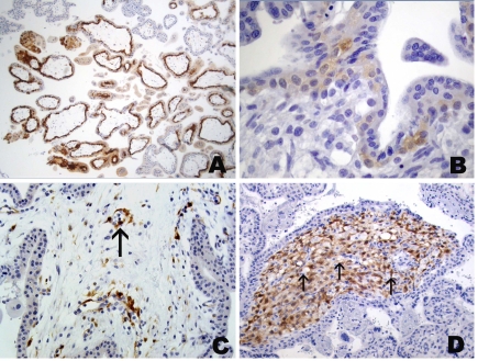
Figure 6.
Immunohistochemical staining of placenta with mAb ES121 to NY-ESO-1. Immunoreactivity in a week 6 placenta with predominant staining of cytotrophoblast, weaker immunoreactivity in syncytiotrophoblast and strong staining in several cell columns (A). (B) More commonly seen, very weak reactivity of villous cytotrophoblast in a week 10 placenta. (C) Focal immunopositive cells in villous stroma of week 6 placenta consisting of endothelia (arrow) and fibroblasts. (D) Homogeneous immunopositivity in villous stroma of week 18 placenta; note negative vacuolated Hofbauer cells (arrows).

