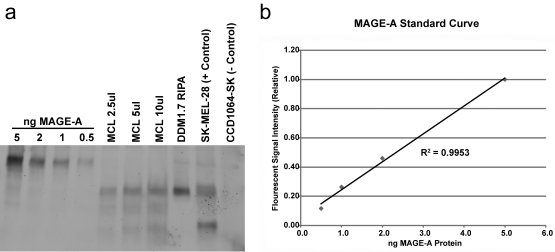
Figure 4.
MAGE-A protein is present in DDM1.7 cell lysate. (a) Western blot using the pan-MAGE-A monoclonal antibody 6C1 (reacts with MAGE-A1, -A2, -A3, -A4, -A6, -A10, and -A12). Samples analyzed: “ng MAGE-A” indicates the amount of MAGE-A fusion protein loaded in each lane, MCL corresponds to melanoma cell lysate, DDM1.7 RIPA refers to whole cell lysate made with RIPA buffer, SK-MEL-28 is a positive control cell lysate, CCD1064-SK is a negative control cell lysate. (b) Graph showing the MAGE-A standard curve made by measuring the fluorescent intensity of MAGE-A fusion protein in the Western blot shown above.

