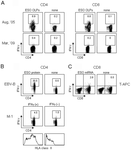
Figure 3.
IFNγ secretion assays. MACS beads-purified CD4 and CD8 T cells (2 x 10⁶) from PBMCs were cultured with irradiated (40 Gy) autologous CD4- and CD8-depleted PBMCs (2 x 10⁶) as APCs in the presence of 28 overlapping 18-mer peptides and a 30-mer C-terminal peptide (OLPs) spanning the entire NY-ESO-1 protein (1 µg of each peptide/ml) in 24-well culture plates for 12 days. (A) IFNγ secretion by CD4 and CD8 T cells (1 x 10⁵) was assayed against PFA-treated CD4- and CD8-depleted PBMCs (1 x 10⁵) pre-pulsed with NY-ESO-1 OLPs for 30 min. (B and C) CD4 and CD8 T cells obtained in August 2005 were used on the twenty-sixth day following two stimulations. (B) IFNγ secretion by CD4 T cells (1 x 10⁵) was assayed against the patient's EBV-transformed B cells (1 x 10⁵) pretreated with NY-ESO-1 protein (20 µg/ml) for 24 h, and NY-ESO-1-expressing melanoma (M-1) cells (1 x 10⁵) pretreated with IFNγ (100 U/ml) for 48 h by stimulation for 4 h. HLA class II expression on M-1 cells after IFNγ treatment is also shown. (C) IFNγ secretion by CD8 T cells (1 x 10⁵) was assayed against the patient's PHA-stimulated CD4 T cells (T-APC) (1 x 10⁵) transfected with NY-ESO-1 mRNA (20 µg).

