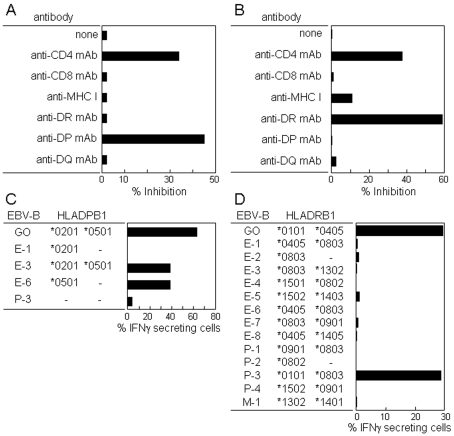
Figure 5.
Restriction molecules involved in the CD4 T cell recognition of NY-ESO-1. CD4 T cell recognition of peptide 16 (aa 91-108) (A and C) and peptide 21 (aa 121-138) (B and D) was analyzed by antibody blocking (A and B) and using various EBV-B cells as APCs (C and D). CD4 T cells cultured with irradiated (40 Gy) autologous CD4- and CD8-depleted PBMCs in the presence of a mixture of OLPs for 14 days, as described in the legend of Figure 4, were assayed for IFNγ secretion against PFA-treated autologous CD4- and CD8-depleted PBMCs pre-pulsed with peptide 16 (aa 91-108) (A) and peptide 21 (aa 121-138) (B) in the presence of various mAbs (2 µg/ml) during the assay, and against PFA-treated EBV-B cells as APCs pre-pulsed with peptide 16 (aa 91-108) (1 µg/ml) (C) and peptide 21 (aa 121-138) (1 µg/ml) (D) for which HLA genotypes have been determined. IFNγ production was determined by ELISA in A and B using the supernatant after culture for 18 h and by an IFNγ secretion assay in C and D after stimulation for 4 h.

