
Figure 1.
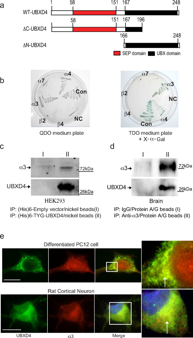
UBXD4 interacts with α3* and α4* nAChRs. A yeast two-hybrid screen showed that the large intracellular loop of the α3 nAChR subunit interacts with a novel UBX-containing protein named UBXD4. a, Schematic diagrams illustrating the structural location of the UBX and SEP domains in WT-UBXD4 (top panel). Middle and bottom panels represent the original clone 42 found in Y2H (ΔC-UBXD4, middle) and the generated ΔN-UBXD4 clone lacking the SEP domain (bottom). b, Interaction was verified with cotransformation of α3 and UBXD4 into the Y187 yeast strain. Further experiments in yeast showed that UBXD4 interacts with α4 but not with the α7, β2, and β4 nAChR subunits demonstrating that UBXD4 is a selective partner for the α3 and α4 subunits. Left panel, Quadruple dropout media (−Leu/−His/−Trp/−Ade). Right panel, Triple dropout media (−Leu/−His/−Trp) containing X-α-gal for α-galactosidase activity; CON, positive control; NC, negative control. c, In vitro experiments with charged Ni+ columns showed that the α3 subunit can bind to (His)6-TYG-tagged UBXD4 protein expressed in HEK293 cells. d, UBXD4 interacts with native α3 nAChR subunits from mouse brain. PFC lysates were incubated either with rabbit IgG (I, control) or the anti-α3 antibody (II) immobilized on protein A/G. Eluted proteins were subjected to SDS-PAGE and immunoblotted with the anti-α3 and anti-UBXD4 antibodies. e, Differentiated PC12 cells endogenously expressing α3-containing nAChRs (top) or rat cortical neurons (bottom) were cultured on coverslips, and processed for immunocytochemistry. Staining of the permeabilized cells for UBXD4 (green) and α3 (red) demonstrated partial overlapping of UBXD4 and α3 in the perinuclear region of the cells, as indicated by the white arrows in the magnified inset. Nonspecific binding or staining of the nucleoplasm in PC12 cells was observed by omitting the primary antibody during the incubation process (Fujiwara et al., 2006). Confocal scale bar, 10 μm.