
Figure 8.
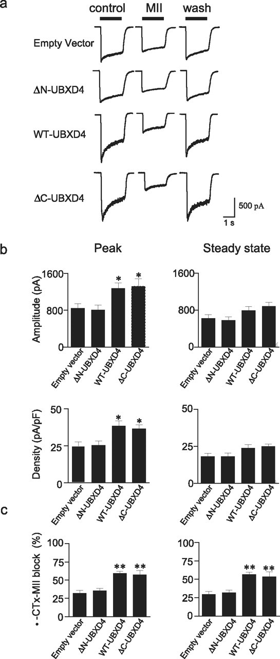
Functional regulation of α3* nAChRs by UBXD4. a, Currents elicited by 300 μm ACh were measured before, during, and after a 5 min exposure to 100 nm α-CTx-MII in dPC12 cells stably transfected with empty vector, ΔN-UBXD4, WT-UBXD4, or ΔC-UBXD4. b shows peak (left panels) and steady-state (right panels) current amplitude and density in the various cell lines described in a. Overexpression of WT-UBXD4 and ΔC-UBXD4 produced a significant increase (p < 0.05) in peak amplitude and current density compared with empty vector or ΔN-UBXD4. c, Block by α-CTx-MII of peak (left panel) and steady-state (right panel) α3* nAChR currents. The α-CTx-MII-sensitive component was greater in cells overexpressing WT-UBXD4 and ΔC-UBXD4 than in cells expressing the corresponding empty vector or ΔN-UBXD4 (p < 0.01). Values represent mean ± SEM values from cells expressing empty vector (n = 16), ΔN-UBXD4 (n = 12), WT-UBXD4 (n = 34), and/or ΔC-UBXD4 (n = 8), respectively.