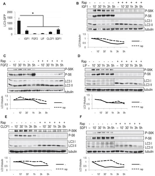
Figure 4.
Cytokines identified in the screen suppress autophagy independently of mTORC1 activity. A, Quantification of autophagy in H4 LC3-GFP cells grown in serum-free medium supplemented with indicated cytokines for 24h. All error bars are s.e.m. * p<0.05 n≥8 B–F, Cytokines are able to suppress autophagy in the absence and presence of rapamycin. H4 cells were grown in serum-free medium, followed by addition of 100 ng/mL IGF1 (B), 50 ng/mL FGF2 (C), 50 ng/mL LIF (D) or 50 ng/mL CLCF1 (F) and 10 μg/mL E64d. Where indicated, cells were pre-treated with 50 nM rapamycin 1 hour prior to the addition of cytokines. Levels of autophagy were assessed by western blot using antibody against LC3; mTORC1 activity was evaluated with antibodies against phospho-S6 (Ser235/236, P-S6) and phospho-S6 kinase (Thr389, P-S6K). Quantification of LC3 II/tubulin ratio is shown.

