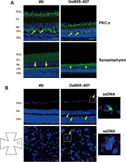
Figure 4.
Degeneration of outer plexiform layer (OPL) and outer nuclear layer at the peripheral retina of Del605–607 mouse. (A) Immunostaining of the retina sections with tyrosine hydroxylase (red) and PKC α (green), a specific maker for dopaminergic amacrine cells and rod bipolar cells, respectively (upper panel). Cell loss and size reduction of bipolar cells was observed. Scale bar, 20 µm. Immunostaining of the retina sections with synaptophysin, a specific marker for neuronal presynaptic vesicles (lower panel). Synapse disruption (arrow) was observed in the OPL of Del605–607 mouse peripheral retina. Scale bar, 50 µm. (B) Immunostaining of cryosection and flat mount retina with specific apoptosis marker, ssDNA. Apoptosis cells were observed in the all cell layers in the peripheral retina of Del605–607 mice. Scale bar, 50 µm.

