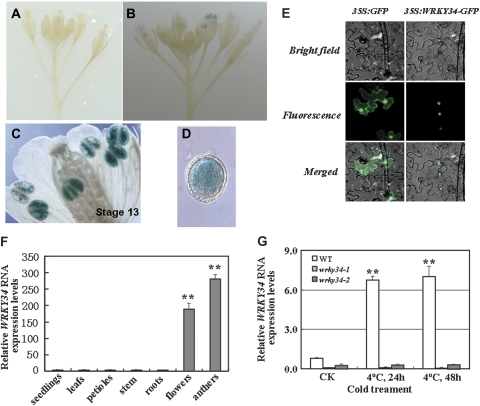
Fig. 1.
Expression patterns and subcellular localization of WRKY34. (A) A negative control non-transgenic wild-type (WT) inflorescence, showing no GUS activity. (B) A transgenic inflorescence, showing GUS staining in the anthers. (C) Transgenic mature pollen grains with GUS staining at the floral developmental stage 13. (D) Transgenic mature pollen grains with GUS staining. (E) The subcellular localization of the WRKY34–GFP fusion protein in Nicotiana benthamiana epidermal cells. Images in the left column show the control plasmid expressing only the green fluorescent protein (GFP) and those in the right column show the WRKY34–GFP fusion protein expressed in N. benthamiana epidermal cells. The cells were examined with brightfield (top) and UV fluorescence (middle) microscopy, and as a merged image (bottom) showing either the diffused (control plasmid) or the nuclear localization of the proteins. (F) Quantitative RT-PCR comparison of the WRKY34 mRNA expression levels in different tissues from the WT plants grown at 22 °C. (G) Quantitative RT-PCR comparison of WRKY34 RNA expression levels in cold-treated (4 °C) and untreated (22 °C) mature pollen of the WT and wrky34 mutant. After cold treatment, WRKY34 RNA expression levels significantly increased in the WT, and showed no distinct change in the wrky34 mutants. Error bars indicate standard deviations of three independent biological samples. Differences between the untreated and treated plants with cold stress are significant at P <0.01 (**).

