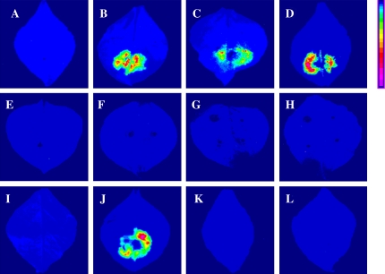
Fig. 5.
Firefly luciferase complementation imaging assay of OsARF–OsIAA protein interactions in N. benthamiana leaves. Leaves were co-infiltrated with Agrobacterium containing the following vector pairs: (A) negative control, OsARF16-NLuc/CLuc; (B) positive control, SGT1a-NLuc/CLuc-RAR1; (C) OsIAA1-NLuc/OsIAA1-CLuc; (D) OsIAA12-NLuc/OsIAA12-CLuc; (E) OsARF1- NLuc/OsIAA1-CLuc; (F) OsARF1-NLuc/OsIAA8-CLuc; (G) OsARF1-NLuc/ OsIAA12-CLuc; (H) OsARF1-NLuc/OsIAA13-CLuc; (I) OsARF16-NLuc/OsIAA1- CLuc; (J) OsARF16-NLuc/OsIAA8-CLuc; (K) OsARF16-NLuc/OsIAA12-CLuc; (L) OsARF16-NLuc/OsIAA13-CLuc. Pseudocolour bar, right, shows the range of luminescence intensity from weak to strong. Images were collected 3 d after infiltration. The results were tested independently in triplicate. (This figure is available in colour at JXB online.)

