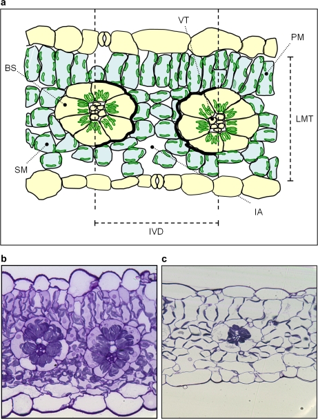
Fig. 5.
(a) Diagram representing regions of interest (ROIs) in cross-sections of Flaveria bidentis leaves. ROIs include bundle sheath cells (BS), palisade mesophyll cells (PM), spongy mesophyll cells (SM), and total vascular tissue (VT) which were manually outlined and measured to give area and perimeter data. Interveinal distances (IVD) were measured between two adjacent vascular bundles. Leaf mesophyll thickness (LMT) was measured between epidermal layers at four points in each cross-section. Intercellular airspaces (IA) within the interveinal distance were individually outlined and combined to give a total length of mesophyll cells exposed to intercellular airspace (Sm). Black stripes represent the perimeter of the bundle sheath tissue within the interveinal zone (Sb). Representative light microscope images (×400 magnification) of leaf sections of plants grown at 150 μmol quanta m−2 s−1 (b) and 500 μmol quanta m−2 s−1 (c). (This figure is available in colour at JXB online.)

