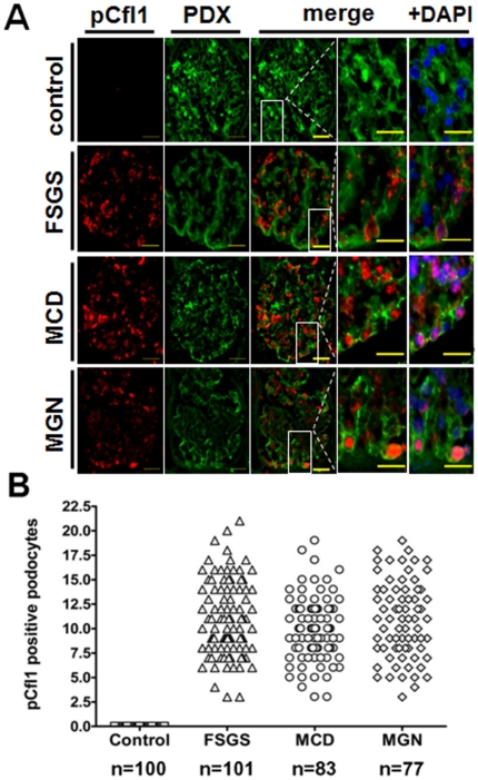
Figure 5. Phosphorylated cofilin-1 was increased in podocyte nuclei of patients suffering from FSGS, MCD and MGN.
Glomeruli recovered from patients with glomerular diseases, FSGS, MCD and MGN, were probed with the rabbit primary antibody to p-cofilin and goat primary antibody to podocalyxin (PDX) and visualized using an Alexa488 labelled antibody raised against goat (green) and a Cy3-labelled secondary antibody against rabbit (red). Podocytes in these diseased glomerular tissues demonstrated an increased incidence of phosphorylated cofilin-1 in their nuclei (merged images ±DAPI). In contrast p-cofilin of normal control patients revealed no glomerular staining indicating that the cofilin-1 expressed in the glomerulus under normal conditions is active and under disease-conditions is inactivated.

