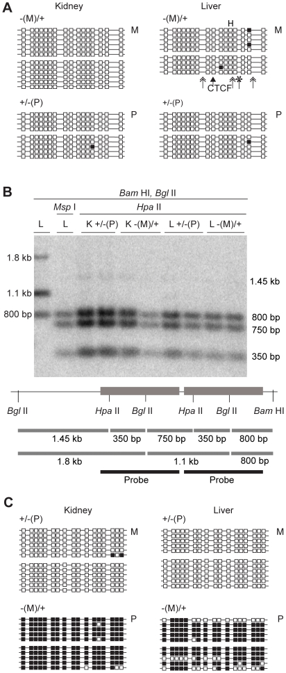
Figure 4. DNA methylation of the (mChβGI)2 in 18.5 dpc fetuses.
(A) Bisulfite sequencing was performed to analyze CpG methylation of the (mChβGI)2 using genomic DNA from 18.5 dpc fetuses. Genotypes are indicated on top. Maternal (M) or paternal (P) transmission of the allele is indicated on the right. Unmethylated CpGs (white squares) and methylated CpGs (black squares) are shown along independent chromosomes (horizontal lines). Two siblings were assessed in each case, separated by space between groups of chromosomes. Simple arrow indicates the CTCF site. Double arrows and asterisk indicate the positions of the USF1 and VEZF1 deletions. (B) Southern blot hybridization results in kidneys (K) and livers (L) after paternal and maternal transmission. The (ChβGI) sequence was used as a probe. The two diagnostic HpaII/MspI sites and the BamHI and BglII restriction sites are indicated. (C) Bisulfite sequencing of the ICR sequences from the same samples as in (A).

