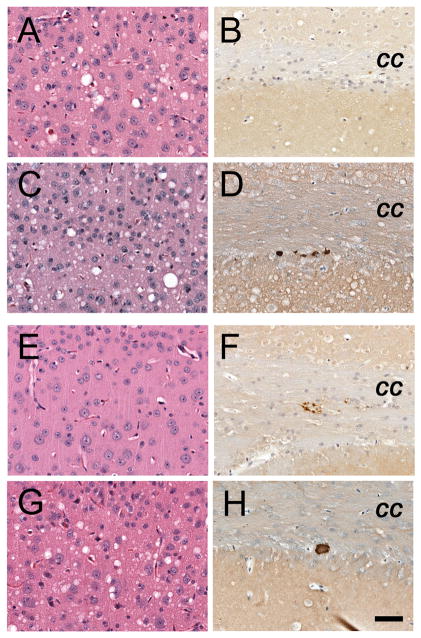
Fig. 5.
Neuropathology resulting from serial passage of brain from vCJD-inoculated mice with either type 1 PrPSc (A–D) or type 2 PrPSc (E–H), in Tg1014 mice. For the type 1 strain, neuropathologic changes upon second (A, B) and third (C, D) passages were similar to first passage (Fig. 3E–G): moderate vacuolation (A, C) and few amyloid deposits (B, D) were observed. For type 2 PrPSc, second passage (E, F) showed little vacuolation (E) similar to first passage (Fig. 3I), but few amyloid plaques and sparse PrPSc deposits (F). Upon third passage of type 2 PrPSc (G, H), neuropathology resembled that of type 1 PrPSc with numerous vacuoles (G) and very few amyloid plaques (H). Sections stained with H & E (left column), and PrP immunohistochemistry (right column). Bar in panel H represents 50 μm and applies to all panels.

