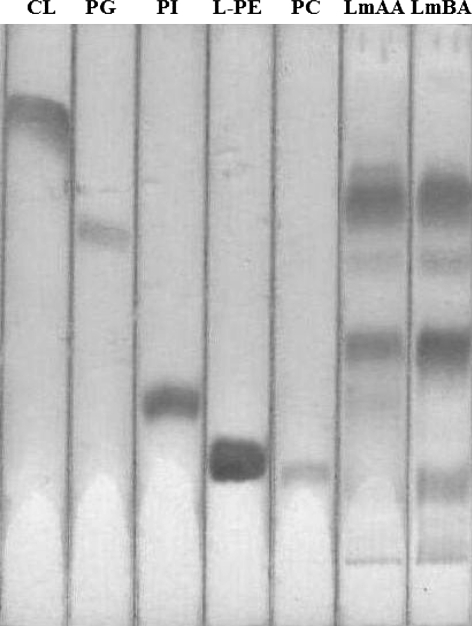
Fig. 3.
Thin-layer chromatography separation of individual L. monocytogenes phospholipids derived from cells grown in presence of acetic LmAA or benzoic acid LmBA (6th and 7th zone, respectively, from the left). The diagram shows these standards from the left to right: CL cardiolipin, PG phosphatidylglycerol, PI phosphatidylinositol, L-PE lyso-phosphatidylethanolamine and PC phosphatidylcholine. The plate was developed using the solvent system chloroform/methanol/acetic acid/water [50/25/6/2, vol/vol/vol/vol]. Visualisation: after spraying with molybdenum blue reagent. L. monocytogenes cells contained, from top to bottom: CL, PG, PhAL (phosphoaminolipid), PI, and phosphocomponent A

