Figure 5.

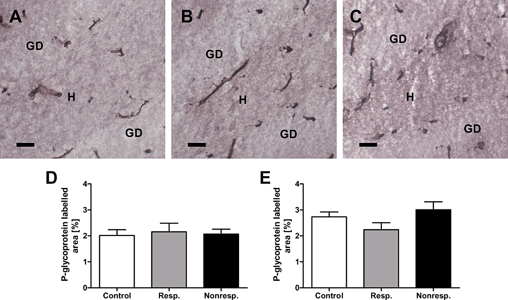
Celecoxib treatment of responders (n = 4) and non-responders (n = 4) decreases P-glycoprotein expression to control level. (A) Representative immunostaining of P-glycoprotein in the hippocampal dentate hilus [H] of a non-epileptic control rat (DG, dentate gyrus). (B) P-glycoprotein immunostaining in the hippocampal dentate hilus of an epileptic rat responding to phenobarbital (responder) during the first and second treatment phase. Note that brain tissue samples have been taken following celecoxib pretreatment and the second phenobarbital treatment phase. (C) P-glycoprotein immunostaining in the hippocampal hilus of an epileptic rat that was classified as a non-responder during the first phenobarbital treatment phase. Note that this rat responded to phenobarbital following celecoxib pretreatment, that is, during the second phenobarbital treatment phase and that brain tissue samples have been taken following celecoxib pretreatment and the second phenobarbital treatment phase. P-glycoprotein immunoreactivity in capillaries proved to be comparable between the three groups of rats. Quantitative analysis of P-glycoprotein immunostaining in the hippocampal dentate hilus (D) and the parietal cortex (E). Note that celecoxib treatment reduced P-glycoprotein levels to control levels in responders as well as non-responders. Data are given as mean ± SEM. Scale bar = 100 µm.
