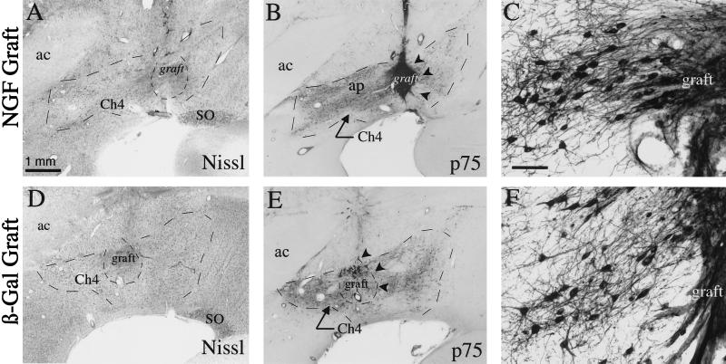
Figure 1.
NGF-secreting and control grafts within Ch4i. (A) Thionin-stained coronal section showing NGF-secreting cell graft within the Ch4i. The graft is well integrated within host tissue. (B) p75 immunolabeled section adjacent to A reveals dense graft penetration by cholinergic axons. (C) Higher magnification of p75 labeled cholinergic neurons adjacent to an NGF-secreting graft demonstrates fiber growth directed toward the graft. (D–F) Comparable thionin and p75 immunolabeled sections from a control aged monkey that received β-galactosidase-expressing fibroblasts. Graft survival is comparable to that of NGF grafts, but fewer axons penetrate the grafts. ac, anterior commissure; ap, Ansa peduncularis; SO, supraoptic nucleus. (Scale bar in A = 1 mm and applies to A, B, D, E; bar in C = 100 μm and applies to C and F.)

